
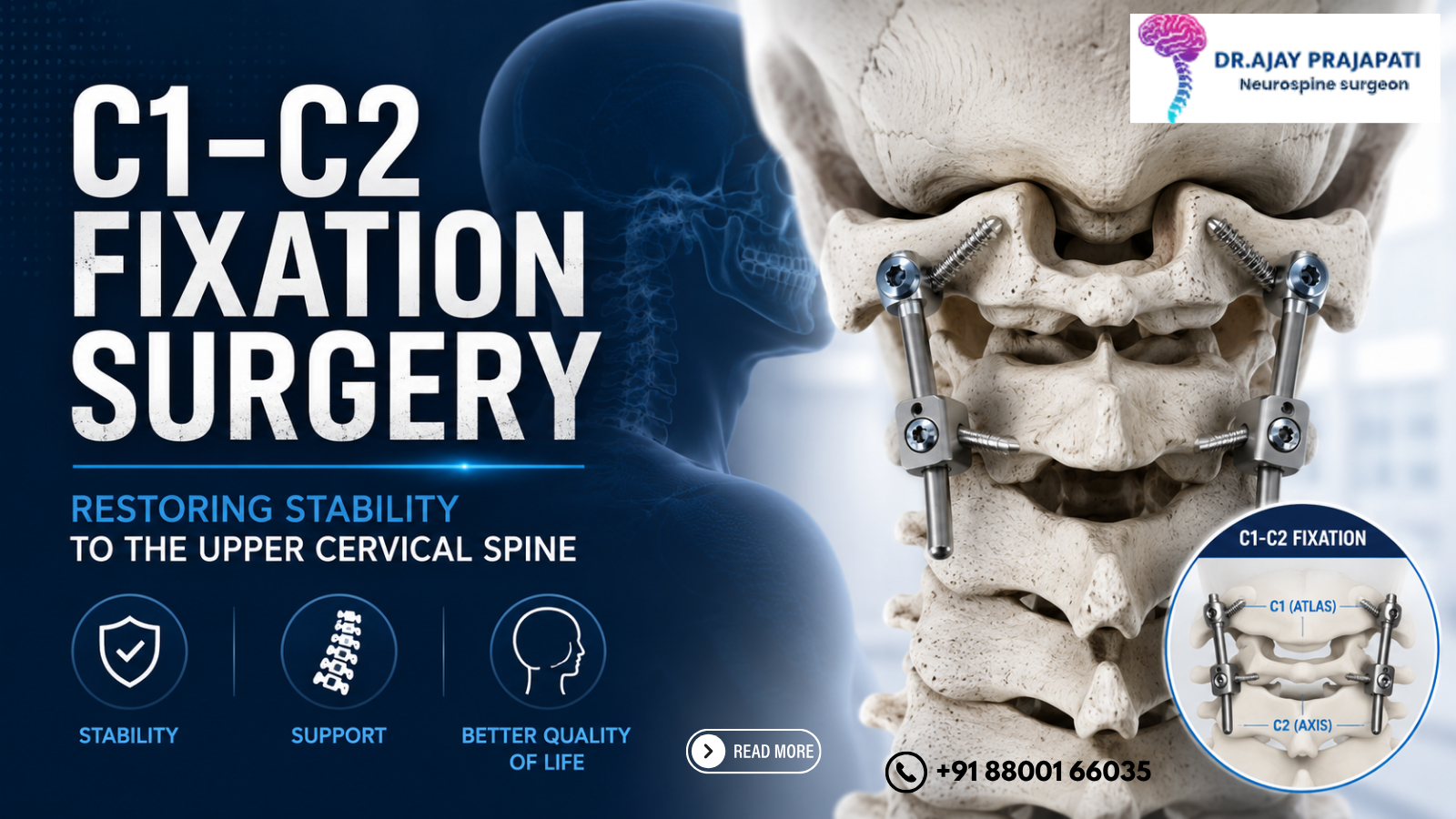
C1-C2 Fixation Surgery: Restoring Stability to the Upper Cervical Spine
At the very top of the spine, just beneath the skull, sit two of the most critically important vertebrae in the entire human body — C1 (the atlas) and C2 (the axis). Together, they form the craniocervical junction: a uniquely complex anatomical region responsible for supporting the weight of the skull, protecting the brainstem and upper spinal cord, and enabling the head to rotate, nod, and tilt through its remarkable range of motion.
When this junction becomes unstable — through trauma, disease, congenital abnormality, or degeneration — the consequences can be catastrophic. An unstable C1-C2 joint places the brainstem and upper cervical spinal cord under constant, potentially fatal mechanical stress. The solution, when surgical, is C1-C2 fixation: a complex but highly effective procedure that permanently stabilises the craniocervical junction and protects the neural structures within.
For patients across Delhi-NCR navigating a diagnosis of C1-C2 instability, access to the best spine surgeon in Noida with dedicated craniocervical expertise is not just important — it is essential. This blog explains what C1-C2 fixation surgery involves, who needs it, how it is performed, and what recovery looks like.
Understanding C1 and C2: Anatomy and Function
C1 — The Atlas
C1, named after the Greek Titan who carried the world on his shoulders, is a ring-shaped vertebra with no vertebral body. It sits atop C2 and cradles the base of the skull (the occipital condyles) in its lateral masses. C1 is responsible for approximately 50% of the head's nodding (flexion-extension) movement.
C2 — The Axis
C2 has a unique bony protrusion called the dens (odontoid process) — a peg-like projection that rises upward from its body and fits into a groove on the inner surface of C1's anterior arch. This articulation forms the atlantoaxial joint, which is responsible for approximately 50% of the head's rotational movement — allowing us to turn our heads left and right.
The dens is held in position by the transverse ligament — a powerful horizontal band that prevents the dens from pressing backward into the spinal cord. When this ligament is torn, stretched, or compromised, or when the dens itself is fractured, the entire C1-C2 articulation becomes dangerously unstable.
What Causes C1-C2 Instability?
C1-C2 instability is a serious condition with several distinct causes:
1. Odontoid Fractures (Dens Fractures)
The dens is one of the most commonly fractured bones in the cervical spine, particularly in high-velocity road accidents and falls in elderly patients (where even minor falls can cause dens fractures due to osteoporosis). Type II odontoid fractures — at the base of the dens — are the most common and most likely to require surgical fixation due to their poor healing potential.
2. Atlantoaxial Instability from Ligamentous Injury
High-energy trauma — road accidents, diving injuries, contact sports — can rupture the transverse ligament, eliminating the primary restraint holding the dens away from the spinal cord. This is an immediate surgical emergency.
3. Rheumatoid Arthritis
One of the most common systemic causes of C1-C2 instability. Rheumatoid arthritis causes inflammatory erosion of the synovial joints and ligaments of the craniocervical junction, allowing progressive anterior atlantoaxial subluxation (the atlas slides forward relative to the axis). Approximately 25–40% of patients with longstanding rheumatoid arthritis develop some degree of cervical spine involvement.
4. Congenital Anomalies
Conditions such as os odontoideum (failure of the dens to fuse with the C2 body during development), Down syndrome (associated with atlantoaxial instability due to ligamentous laxity), and Klippel-Feil syndrome predispose patients to craniocervical junction instability from birth.
5. Craniovertebral Junction Anomalies
Basilar invagination — where the top of C2 migrates upward into the skull base — and Chiari malformation with associated instability are increasingly recognised causes of upper cervical instability requiring surgical correction.
6. Tumours and Infections
Primary bone tumours, metastatic cancer deposits, and spinal infections (tuberculosis of the spine — Pott's disease — is particularly relevant in India) can destroy the bony integrity of C1 or C2, leading to instability.
7. Degenerative Disease
Though less common than at lower cervical levels, severe degeneration of the C1-C2 facet joints can cause pain and progressive instability, particularly in older patients.
Symptoms of C1-C2 Instability
The clinical presentation of C1-C2 instability varies from subtle to catastrophic, depending on the degree of instability and the extent of neural compression:
Pain
Severe neck pain — often the first and most prominent symptom
Occipital headache — pain at the back of the head, often radiating to the top of the skull; caused by compression of the C2 nerve root (greater occipital nerve)
Pain worsening with head movement, particularly rotation and flexion
Neurological Symptoms
Myelopathy — weakness, stiffness, and clumsiness of the hands and legs; difficulty with fine motor tasks; unsteady gait
Sensory disturbances — numbness or tingling in the arms, legs, or trunk
Lhermitte's sign — an electric shock sensation running down the spine when the neck is flexed
Hyperreflexia — exaggerated deep tendon reflexes indicating upper motor neuron compression
Brainstem Compression Symptoms
When instability is severe enough to compress the brainstem:
Dysphagia — difficulty swallowing
Dysarthria — slurred speech
Nystagmus — involuntary eye movements
Breathing difficulties — in the most severe cases, brainstem compression can affect the respiratory centres and be life-threatening
Drop attacks — sudden falls without loss of consciousness
In Trauma Settings
Acute high-energy injury causing C1-C2 instability may present with immediate quadriplegia, respiratory failure, or sudden death — underscoring the critical importance of proper spinal immobilisation at the accident scene.
Diagnosis: Imaging Is Everything
Accurate, detailed imaging is the foundation of C1-C2 instability diagnosis and surgical planning:
Plain X-Rays (Flexion-Extension Views)
Dynamic flexion-extension X-rays of the cervical spine can demonstrate instability that is not apparent on static views — the atlantodens interval (ADI) widens beyond 3mm in adults (5mm in children) in atlantoaxial instability.
CT Scan of the Cervical Spine
High-resolution CT with 3D reconstruction provides exquisite detail of bony anatomy — fracture pattern, degree of subluxation, bone quality, and the dimensions of the C1 lateral masses and C2 pedicles that will receive the fixation screws. CT is mandatory for surgical planning.
MRI of the Cervical Spine and Craniocervical Junction
MRI evaluates the spinal cord, brainstem, transverse ligament, and surrounding soft tissues — identifying the degree of neural compression, cord signal change (indicating established myelopathy), and associated disc or ligamentous injury.
CT Angiography (CTA)
The vertebral arteries run through the transverse foramina of C2 and are at risk during C2 pedicle screw placement. CTA preoperatively maps the course and dominance of the vertebral arteries, allowing the surgeon to avoid catastrophic vascular injury.
C1-C2 Fixation Surgery: The Procedure Explained
C1-C2 fixation aims to achieve rigid mechanical stabilisation of the atlantoaxial joint, decompress any compressed neural structures, and provide an environment for bony fusion — creating a permanent, stable union between C1 and C2.
Positioning and Approach
The patient is positioned prone (face down) on a specialised neurosurgical frame with the head held in a Mayfield head clamp in the optimal position — typically slight extension to reduce anterior subluxation. Intraoperative fluoroscopy or 3D navigation is used continuously throughout the procedure.
A posterior midline incision is made from the occiput (base of skull) to C3, and the muscles are carefully dissected away from the posterior elements of C1 and C2.
The Goel-Harms Technique: The Gold Standard
The most widely used technique for posterior C1-C2 fixation is the Goel-Harms technique, which uses:
C1 lateral mass screws — inserted into the thick lateral masses of the atlas on each side; these are the anchor points on the C1 side
C2 pedicle screws (or isthmus/pars screws) — inserted through the pedicles of the axis; the pedicle of C2 is dense, strong bone that provides excellent screw purchase
The C1 and C2 screws on each side are connected by titanium rods, which are locked into position using set screws. This construct creates immediate, rigid stabilisation of the C1-C2 joint — stopping all pathological motion and protecting the spinal cord and brainstem from further injury.
Decompression (When Required)
If the spinal cord or brainstem is being compressed by the dens, by thickened ligaments, or by granulation tissue (particularly in rheumatoid arthritis), decompression is performed before or in addition to fixation:
Transoral odontoidectomy — surgical removal of the dens through the mouth; used when anterior decompression is required that cannot be achieved from behind
Posterior arch resection — removal of the posterior arch of C1 when it is contributing to cord compression from behind
Bone Grafting and Fusion
To achieve permanent bony union between C1 and C2, bone graft material is placed between the decorticated posterior surfaces of C1 and C2. Options include:
Autologous iliac crest bone graft — the patient's own bone from the hip; the gold standard for fusion
Local bone graft — bone shavings from the surgical site
Synthetic bone substitutes — increasingly used to avoid harvest site morbidity
Fusion typically becomes radiographically solid over 3–6 months, at which point the titanium instrumentation is no longer the primary load-bearing structure — the fused bone takes over.
Navigation and Neuromonitoring
Modern C1-C2 fixation is performed with:
Intraoperative CT-based navigation — allows real-time, three-dimensional guidance of screw placement with sub-millimetre accuracy; dramatically reduces the risk of screw malposition and vertebral artery injury
Intraoperative neurophysiological monitoring (IONM) — continuous monitoring of spinal cord function through somatosensory evoked potentials (SSEPs) and motor evoked potentials (MEPs); provides immediate warning if neural function is compromised during surgery
The combination of advanced navigation and continuous neuromonitoring has transformed the safety profile of this technically demanding procedure. The best spine surgeon in Noida performing C1-C2 fixation should have access to both — and the experience to interpret and act on them.
Risks and Complications
C1-C2 fixation is one of the most technically demanding procedures in spinal surgery, and patients should have a thorough understanding of the potential risks:
Vertebral artery injury — the most feared complication; can cause posterior circulation stroke, cerebellar infarction, or death; risk is minimised by preoperative CTA and intraoperative navigation
Spinal cord injury — worsening of existing neurological deficits or new deficits; rare in experienced hands with adequate neuromonitoring
C2 nerve root injury — causes persistent occipital numbness or pain
Screw malposition — may require revision; navigation dramatically reduces this risk
Non-union (pseudarthrosis) — failure of the bone graft to fuse; may require revision surgery
Infection — deep surgical site infection is rare but serious; it requires prolonged antibiotic therapy and sometimes surgical debridement
CSF leak — dural tear with cerebrospinal fluid leakage; usually manageable intraoperatively
Overall, in experienced centres with modern navigation and neuromonitoring, the risk of serious complications is low, and must be weighed against the risk of no surgery, which in most cases of C1-C2 instability means progressive neurological deterioration and potentially fatal brainstem compression.
Recovery After C1-C2 Fixation Surgery
Immediately Post-Operative
Most patients are monitored in the ICU or high-dependency unit for 24–48 hours following surgery. A post-operative CT scan is performed to confirm screw position and alignment. A cervical collar is worn for 6–12 weeks to protect the construct during early healing.
Hospital Stay
Most patients are discharged within 3–5 days after an uncomplicated procedure, provided neurological status is stable or improving.
Neurological Recovery
Neurological recovery following decompression and stabilisation follows a predictable pattern — the most rapid improvement occurs in the first 3–6 months, with continued slower recovery over 12–18 months. Patients with long-standing myelopathy before surgery may have incomplete recovery — underscoring the importance of early surgical intervention before irreversible cord changes develop.
Activity and Rehabilitation
No driving for a minimum of 6 weeks; longer if neurological deficits are present
Physiotherapy begins at approximately 6 weeks — focused on gentle neck mobilisation, strengthening of cervical musculature, and postural rehabilitation
Return to desk work typically at 4–6 weeks; physically demanding work at 3–6 months
Contact sports and high-risk activities are permanently restricted in most patients following C1-C2 fusion, given the loss of rotational movement at this level
Loss of Rotation
The most significant functional consequence of C1-C2 fusion is the permanent loss of approximately 50% of the head's rotational range. This requires adaptation — patients must turn their entire trunk to compensate when looking to the side. Most patients adapt well over time, particularly when the procedure has relieved severe pain and neurological symptoms.
Frequently Asked Questions (FAQs)
Q1. Will I lose neck movement after C1-C2 fixation surgery?
Yes — approximately 50% of rotational head movement is lost, but nodding is largely preserved, and most patients adapt well over time.
Q2. How long does C1-C2 fixation surgery take?
Typically, 2–4 hours depending on complexity, whether decompression is required, and whether navigation is used.
Q3. Is C1-C2 fixation surgery safe?
In experienced hands with modern navigation and neuromonitoring, it is safe — though it carries real risks that must be discussed thoroughly with your surgeon.
Q4. How long before fusion is confirmed after C1-C2 fixation?
Bony fusion is typically confirmed on a CT scan at 3–6 months post-operatively.
Q5. Where can I find an experienced C1-C2 spine surgeon in the NCR region?
Consult the best spine surgeon in Noida with dedicated craniocervical expertise, intraoperative navigation, and neuromonitoring capabilities for this technically demanding procedure.
Conclusion
C1-C2 fixation surgery is one of the most technically demanding and consequential procedures in all of spinal surgery. It addresses instability at the most critical junction in the entire spine — where the brain meets the neck — and when performed correctly, it can be genuinely life-saving and life-transforming.
The anatomy is complex, the surgical risks are real, and the margin for error is slim. But modern advances — intraoperative CT navigation, real-time neuromonitoring, the Goel-Harms technique, and preoperative vertebral artery mapping — have made this procedure far safer than it was even a decade ago.
If you or a loved one has been diagnosed with C1-C2 instability, atlantoaxial subluxation, an odontoid fracture, or craniocervical junction pathology — do not delay seeking specialist evaluation. Connect with the best spine surgeon in Noida for a comprehensive assessment, advanced imaging review, and a clear, expert-guided treatment plan.
Your neck — and everything it protects — deserves nothing less.